All About Computer Training and Learning, Also Sure Shahbaz Khan Sir Materials Sharing Connection. Materials About All Things Here You Can Download, Software Programming Projects ,Notes And Books, And Also Video Tutorials, So Join Us Sure Shahbaz Khan Sir. Any Question And Comments So Please Fill Contact Form In Below Black Button. And My Contact No: 8149155620

August 2016
3D Max (Tutorials) 3D Max (Course) 3D Max (Working) ADCA (Course) Advance Diploma Course Agriculture Alluminium Ambulance Android ( Working ) Android (Tutorials) Android App (Course) Announcement ASP ( Working ) ASP (Course) ASP (Tutorials) ATM ATM and INSURANCE Auto Consultant Auto Parts Autocad (Course) AutoCad (Tutorials) AutoCad (Working) Bags BANK Beauty Parlor Blood Bank Book Download Books and Stationary Breakfast Bricks Bus Timing C Language ( Working ) C Language (Course) C Language (Tutorials) C++ Language ( Working ) C++ Language (Course) C++ Language (Tutorials) Cake Shop Calendar 2019 Car Drivers Car-Rent Caters CCC (Course) Cement Certificate Course Chinese Food CLOTH SECTION Colleges Computer Courses Computer Sales and Service COMPUTER SECTION Computer Training Institute CONSTRUCTION SECTION Corel Draw (Tutorials) Corel Draw (Working) Cosmetics Court Website CSS ( Working ) CSS (Tutorials) CTTC (Course) Current Exam Dates Current Time Table Cyber Cafe Cycle DCA (Course) Designing Digital Printing Diploma Course Dj and Bands Downloads Dreamweaver ( Working ) Dreamweaver (Tutorials) DTH Sales and Service DTP (Course) Dua Eassy Download EDUCATION and LEARNING Electrical Electrician Electronic and Appliance Emergency Facility Family Shop Finance Financial Consultant Fire Brigade Food and Fruit Market Footwear For Born Baby and Kids For Ladies For Mens Forms and Results Four Wheeler Functions Furniture Genreal Store and Masala Gifts Glass Google Sketch Up (Course) Graphics Designer Greeting Article Gym Khana Hall and Lawns Hardware Hardware (Course) Hardware Networking HEALTH and FITNESS Health Wealth Hospital and Clinic Hospitals HOTEL and LODGING How To How To Tutorials HTML ( Working ) HTML (Tutorials) Ice-cream Parlour IFTTT Instagram Instagram Official Insurance ISM Office (Tutorials) Java ( Working ) Java (Course) Java (Tutorials) Java Script ( Working ) Java Script (Tutorials) Jewelry Juice Center Lab Lab Galleries LEGAL and FINANCIAL LIFESTYLE PRODUCT Lodge Malegaon Business Mali and Gavandi Mandap and Stage Decoration Manmad Railway Marbals and Tiles Medical Mess Mobile Sales and Service Money Transfer Motivational Quotes Ms Excel (Tutorials) MS Office (Course) Ms Outlook (Tutorials) Ms Power Point (Tutorials) Ms Word (Tutorials) MS-CIT (Course) Networking (Course) Non-Veg Notes Download Online Admission Online Working Optician Others Our Achievement Our Achivements PageMaker (Tutorials) PageMaker ( Working) Paints Pan-Stall Photo Impact (Tutorials) Photo Impact (Working) Photo Shop (Tutorials) Photo Shop (Working) Photographer PHP ( Working ) PHP (Course) PHP (Tutorials) Pizza and Burger Plumber Police Station Poran Poli PRINTING and GRAPHIC SERVICE Printing Press Programmming Project Download Puja and Diwali Shahitya Pure Veg Python ( Working ) Python (Tutorials) Questions Paper Radium Arts Result Review And Advice Revit (Course) Revit (Tutorials) Revit (Working) Rooms And Hostels Rubber Stamp and Vcard Ruby ( Working ) Ruby (Tutorials) Safety and Security Saloon SAP (Course) Schools Screen Shots Share Brokers Sketch Up (Tutorials) Sketch Up (Working) Software Developer Software Download Steel Study Matrials SunGlasse Suppliment Sure Computers Sweet Homes Tailors Tally (Course) Tayers Thali Three Wheeler Total Students Information TOURS and TRAVEL Toys Tractor Section Travels Tutorial Series Two Wheeler Vb Script ( Working ) Vb Script (Tutorials) Video Editing (Course) Video Intro Video Tutorial Download Video Tutorials VIEW MORE Visual Basic ( Working ) Visual Basic (Course) Visual Basic (Tutorials) Watches Web Designing (Course) Web developer WEDDING and FUNCTION Welfare Computers Whats App Msg Xerox and Binding

August 31, 2016

Amoebic dysentry
Amoebic dysentry

Amoebic dysentry

AMOEBIC DYSENTRY= PATHOGENESIS-     1- it results 4m infection of large intestine by Entamoeba histolytica.  2-caused by ingestion of tetra-nucleated cysts.  3-after ingestion cysts undergo further nuclear division n eight trophozoites r released in terminal ileum. Later they carried to large intestine n produse 'flask shaped' amoebic ulcerations. 4-Incubation period 2-6 wks.     CLINICAL FEATURES= 1-


intesmittent diarrhoea 1-4, foul smelling, loose, watery stools wid mucus n blood.      2-flatulence n abdominal cramping r frequent..3-fever occur sometimes. 4-tender hepatomegaly n tenderness over caecum, ascending colon, left iliac fossa..  COMPLICATIONS= 1-massive haemorrhage. 2-perforation n peritonitis. 3-toxic megacolon. 4-localised granuloma. 5-amoebic liver abscess. 6- amoebic pericarditis. DIAGNOSIS= 1-stool examination show motile trophozoites. 2-sigmoidoscopy, reveal flask shaped ulcers. 3-amoebic serologic tests= indirect haemagglutination test, ELISA,  4- chest radiography 5-ultrasound for amoebic liver abscess. TREATMENTA= 1- metronidazole 800 mg thrice daily for 5 days. Or 2- tinidazole 2 g daily as single dose for 3 days.  3- after treatment furamide 500 mg thrice daily for 10 days. 4- iodoquinol n paromomycin r alternatives. 

August 30, 2016
Atherosclerosis
Atherosclerosis

Atherosclerosis

Defn
a progresiv inflmatory disorder of arterial wall i.e charctrised by focal lipid-rich deposits of atheroma dat remain clinicaly silent until they becom large enough 2 impair arterial perfusion / until ulceration / disruption of lesion results in thrombotic oclusion / embolisation of afectd vessel.


Risk factors

i)major constitutionl risk factors
-age mostly byond 4th d
-sex more in men
-genetic factors
-familial & racial factrs

ii)major acquird risk factrs
-hyperlipidaemia
-HTN
-smoking
-DM

iii)minor risk factrs
-obesity
-exognous harmons lik oral contraceptivs
-endogenous estrogen defi
-physicl inactivity
-stresful lif style
-environmental influencs
-heavy alcohol


pathophysiology


i)early atherosclerosis
fatty streaks @ alterd arterial shear stress -> abnormal endothlial function -> dvlpmnt of inflmn -> inflmatry cels mainly monocyts binds 2 receptors of endothelial cels -> migrate into intima -> take up oxidizd LDL 4m plasma -> lipid laden foam cels or macrophages -> extracelular lipid pool in intima -> foam cels die -> releas of contents -> cytokines, grwth factrs releasd -> smooth muscle cels migration into intima & change 4m contractile 2 a repair phenotype in atempt 2 stabiliz atherosclrotic lesion -> formn of atherosclerotic plaque

ii)Advancd athrsclros
-in establishd atherosclerotic plaque macrophages mediate inflmn & smooth muscl cels promot repair
-if inflmn predominates plaque becmes active/unstabl & may b complicted by ulceration & superaded thrombosis
-cytokines by activtd macrophges & may caus intiml smoth muscl cels ovrlying plaque 2 becom senescent
-thining of fibrous cap
-any breach in intgrity of plaque wil expose contents 2 circulatry bld
-platlet agregation & thrombosis dat extnds in2 athromtous plaque & arterial lumen


Clinicl efects

dpends on siz, typ of artries
1)slow luminl narowing causing ischaemia & atrophy
2)suden luminl oclusn causing infarction necr
3)propagation of plaque by formn of thrombi, emboli
4)formn of aneurysml dilatation & eventual ruptur


#Primary prevention
2 complementary strategies

1)Population strategy
-risk factors in whole population thru diet & lifstyle
-advice on smoking / avg cholestrol / exercise / diet / obesity

2)Targeted strategy
-treat high risk
-consider absolut risk of atheromatous cvs diseas
-antihypertensivs, lipid lowring therapy
-apropriate Rx

#Secondry prevention

in whom evidnc of atheromtous vascular diseas e.g periphral vasc diseas / MI offered a variety of Rx
-energetic corection of risk factors lik smoking, HTN, hypercholstrolmia 

August 29, 2016
AMPUTATION
AMPUTATION

AMPUTATION  

Amputation is a procedure where a part of the limb is removed through one or more bones.
   Amputations of lower limb are performed more commonly than that of upper limb.
 
     *INDICATIONS
1. injury is commonest cause of amputation in developing countries. Common cause in adults (25- 30 yrs)
2. Peripheral vascular diseases including DM - common in elderly (50- 75 yrs)
3. Infections eg. Gas gangrene
4. Tumours
5. Nerve injuries
6. Congenital anomalies
     * TYPES
     A) Guillotine or open amputation
   skin is not closed over wound as wound is not healthy
    Operation done after some period to construct stump:-
  1. Secondary closure
  2. Plastic repair
  3. Revision of stump
  4. Re-amputation

       B) Closed amputation
   Skin is closed primarily
   Surgical Principles:-
   1. Torniquet - except in ischaemic limb.
   2. Ex-sanguination - limb is squeezed with Esmarch bandage before torniquet application.
    Contraindicated in malignancy & infection
   3. Level of amputation :-
 decided on the basis of
    - Disease :- disease for which amputation is indicated. Be conservative with dry gangrene & trauma, but liberal with acute life-threatening infection & malignancy
     - Anatomy :- joint must be saved    
      - Suitability for proper functionig :- sometimes length is compromised for efficient functionig.
    4) Skin flaps :- skin over stump should be mobile & normally sensitive.
     5) Muscles :- muscles are cut distal to level of bone with
   - Myoplasty :- opposite group of muscles sutured together
   - Myodesis :- muscles sutured to end of stump
     6) Nerves :- are gently pulled distally into wound & divided with a sharp knife. Large nerves like sciatic nerve contain large vessel & should be ligated before dividing
      7) Major blood vessel should be isolated & doubly ligated using non-absorbable sutures. Torniquet should be before closure
      8) bone level is decided. Sharp edges of cut bone should be made smooth.
      9) Drain :- corrugated rubber drain is used for 48-72 hours.
     10) After Rx
        - Dressing :- two types of dressings are used 1) Soft
                          2) Rigid
     soft dressing is advantageous for wound healing  & early prosthetic fitting
         - Positing & elevation of stump to prevent contracture & promote healing.
          - Exercises :- for maintaining range of motion of proximal joint
           - Wrapping the stump with crepe-bandage helps in healing, shrinkage & maturation
           - Prmsthetic fitting & gait-training started after 3 months
       *COMPLICATIONS
    1. Haematoma :- due to
- inadequate haemostasis
- loosening of ligature
- inadequate drainage
- it results in delayed wound healing & infection
- Rx aspiration  &  a pressure bandage        
   2. Infection :-
- due to PVD, DM or haematoma
- Rx wth AMA
   3. Skin flap necrosis :-
- indicates insufficient blood
circulation
- avoided by taking care at time of designing
   4. Deformities of joints :-
- improper positionig of amputation stump leads to contracture
- Rx passive stretching exercises
    5. Neuroma
- always forms at the end of but nerve
- adhesion to scar makes it painful
- prevented by dividing the nerves sharply at proximal level & allowing it to retract proximally
- Rx excision at more proximal level
   6. Phantom sensation :-
- sensation of amputed limb being  still present
- most prominent immediately after amputation gradually diminish with time


August 28, 2016
Aphakia
Aphakia

APHAKIA

Aphakia literally means absence of crystalline lens from the eye. However,from the optical point of view,it may considered a condition in which  lens is absent from pupillary area. Aphakia produces high degree of hypermetropia.

CAUSES
1. Congenital absence of lens.
2. Surgical aphakia occuring after removal of lens is commonest presentation.
3. Aphakia due to absorption of lens matter is noticed rarely after trauma in children.
4. Traumatic extrusion of lens from eye.
5. Posterior dislocation of lens in vitreous causes optical aphakia.

OPTICS of APHAKIC EYE
1. Eye becomes highly hypermetropic.
2. Total power of eye is reduced to about +44D from +60D.
3. Anterior focal point  becomes 23.2 mm in front of cornea.
4. Posterior focal point is about 31 mm behind the cornea.
5. There occurs total loss of accomodation.

CLINICAL FEATURES
Symptoms
1. Defective vision. Main symptom in aphakia is marked defective vision for both far and near due to high hypermetropia and absence of accommodation.
2. Erythropsia and cyanopsia. Seeing red and blue images. This occurs due to excessive entry of ultraviolet and infrared rays in absence of crystalline lens.

Signs
1. Limbal scar may be seen in surgical aphakia.
2. Anterior chamber is deeper than normal.
3. Iridodonesis. Tremulousness of iris can be demonstrated.
4. Pupil is jet black in colour.
5. Purkinje's image test shows only two images.
6. Fundoscopy examination shows hypermetropic small disc.
7. Retinoscopy reveals high hypermetropia.

Treatment
Modalities for correcting aphakia include..
1. Spectacles prescription has been the most commonly employed method of correcting aphakia.
Advantages- it is cheap,easy and safe method.
Disadvantage- 1. Image is magnified by 30 percent, so not useful in unilateral aphakia.
2. Problem of spherical and chromatic aberrations of thick lenses.
3. Field of vision is limited
4. Prismatic effect of thick glasses
5. Roving ring scotoma
6. Cosmetic blemish.

2. Contact lenses.
Advantages-
1. Less magnification of image.
2. Elimination of aberrations and prismatic effect of thick glasses.
3. Wider and better field of vision.
4. Cosmetically more acceptable.
5. Better suited for uniocular aphakia
Disadvantages
1. More cost
2. Cumbersome to wear.
3. Corneal complications.

3. Intraocular lens implantation. It is commonest modality being employed.

4. Refractive corneal surgery.
1. Keratophakia.
2. Epikeratophakia
3. Hyperopic lasik.

August 27, 2016
Assisted breech
Assisted breech

Assisted breech

ASSISTED Breech delivery :carried by skilled obs  principles :never to rush never pull from bellow  keep fetus with back anterior
STEPS pt is brought to table when anterior buttock and fetal anus r visible  she is placed lithotomy  position or tilted  Antiseptic cleaning  pudendal block is done episiotomy done in all case Patient is encouraged to bear down for easy descent  No touch of fetus policy is used until buttocks delivered with legs flexed
Soon after trunk upto umbilicus born following done  extend leg Pull cord down and nobilised to one side Rotate trunk to bring it anterior BABY is wraped with towel
delivery of arms :should not be extension of arms Arms are delivered one after other only when one axilla is visible by hooking down elbow
HEAD DELIVERY crucial stage  various methods BURNS MARSHALL METHOD  FORCEP delivery method
process baby allowed to hang by  own weight  Supra pubic pressure by hand in downward backward direction  when neck is visible baby is grasped traction is done
Resuscitation of baby 

August 26, 2016
Asthama
Asthama

Asthama

DEF#
chronic imflamatory disease of airway charcterised by an increased responsiveness of tracheobronchial tree to variety
of stimuli.
         Manifest as paroxysm of dyspnoea,cough,wheezing as result of musclespasm,mucosal oedema,&bronchial secreations.
CLASSIFICATION#
1.Erly onset asthama(atopic,allergic,extrinsic)
2.Late onset asthama(non-atopic,intrinsic,idiosyncratic)
ETIOPATHOGENESIS#
2major factors.
1.Bröchial hyperresponsiveness
2.Inflamatory reaction in bronchial wall
3.Roll of allergans
PATHOGENESIS_
cell                 triggers
[                       [
Mediators___>bronchial
[             hyperresponse
]                      |
Inflamation         |
       |               |
       |_Symptoms__|            
CELLS INVOLVED inreleas mediator
¤Mast cells¤macrophages
¤eosinophil¤lymphocytes
MEDIATORS
¤histamine¤prostaglandins(d2,f2,E2)¤thrombaxane¤PAF¤bradykinin
¤leucotrienes(B4,C4,D4,E4)¤subP
¤adenosine¤oxygen radicals
TRIGGERS
¤exercise¤hyperventilation
.¤cold air¤dust & fumes
¤respiratory viral infection
¤emotional stress¤tobaco smoke
¤aspirin,beta blockers
CLINICAL FEATURES
discussed under 3 headings
1.EPISODIC ASTHAMA
¤occurs in episodes with asymptomatic period between
¤spontaneos in onset triggered
by allergans,exerecise,viral infe
2.SEVERE ACUTE ASTHAMA
also called
status asthamaticus
symptoms persist instid of initial
Rx¤severe dyspnoea,unproductiv
cough
¤Signs-sweating,central cyanosis
tachycardia,pulsus paradoxus
3.CRONIC ASTHAMA
chronic cough with mucoid sputum,¤episodes of cough&wheeze during night
PHYSICAL SIGN IN CHEST
seen onely during attack
1.Tachycardia use of accesory
muscles of respiration
2.Percussion,Hyper-resonant
3.Breath sonds-vesicular
4.Rhonchi&wheeze
5.Silent chest airflow insufficient
to produce rhonchi
INVESTIGATIONs
1.Chest radiography
in attck lungs r hyperinflated
complications like pigeon chest,
lobar/segmental collapse may occur
2.Pulmonary function tests
forced expiratory volume in 1min
[FEV1] vital capacity[VC],peak
expiratory flo rate[PEF]
3.Arterial Blood Gas analysis
it shows hypoxia&hypocarbia
4.Skin hypersensitivity test for
allergans & there avoidance
5.Blood& sputum for eosinophilia
#MANAGEMENT#
3 broad headings
A].Avoidance of allergans
B] .Desensitisation imunotherpy
C].Drugs therapy

A].Avoidance of allergans
it is possible when there is single allergan
.B].Desensitisation
repeated S.C inj of extract of
allergans
.C].Drugs
seven catagories of drugs r use
1}.BETA-ADRENERGIC AGONISTS
b1=heart b2=bronchial smoothmu
¤adrenaline_dose0.3-0.5ml of1:1000 sol route-sc ripeated at
interval of 20minutes
¤salbutamol
dose2-4mg tds oraly/2puff 100ug
¤salmeterol
2puffs of25 ug each2/3timeday
¤formeterol
2puffs of6 ug each1/3times day
2}.METHYL XANTHINES
¤theophylline
dose:100-200mg tds 300mg bds
or450-600mg od
¤aminophylline
dose:loading 5mg/kg slowly ouer
20minute.Maitenance:0.5mg/kg/hr
3}CORTICOSTEROIDS
¤hydrocortisone
dose loading-4mg/kg iv followed
by2-3mg 6 hourly
¤prednisolone
40-60mg oraly single mornig dose
¤beclomethasone (200ug) budesonide(200ug),
4}CHROMONES
¤nedocromil sodium
route-inhalation 4mg2/4time
5}.ANTICHOLINERGICS
¤ipratropium bromide
2puffs of20ug each time 4/day
6}LEUKOTRIENE INHIBITORS
¤zafirlukast 20mg 4times a day
¤montelukast10mg once day 

August 25, 2016
Anc antenatal care
Anc antenatal care


Anc antenatal care

Systematic supervision (examination and care ) of a women during pregnacy is called antenatal care
anc comprie of

careful history taking and examination (general and obstretical)
advice given to pregnant women

procedure at first visit


history taking

gravida -denotes pregnant state both present and past irrespective of period of gestation
parity -denotes previous pregnancy beyond the period of viability

EDD

naegeles formula -by adding 9 calender months and 7 days to first day of the last normal (28 days) period

general examination

built, nutrition, hight, weighit, pallor, laundice, tongue, gums, teeth, oedema of legs
bp -disappeasence of sounds rather than muffling of sound is best representation of distolic pressure during pregnancy
heart lungs liver spleen
breasts

obstetrical examination

abdominal
vaginal -done when pt attends clinic for first time before 12 weeks
ultrasound exam.

routine exam.

1 exam. Of blood
for -Hb , ABO ,Rh grouping and VDRL
screening for blood glucose in selected pt
2 urine
for- proteins sugar and pus cells
clean catch specimen of midstream urine is collected
3 cervical cytology study by papanicolau stain has become a routine in many clinics
special investigation
ultrasound exam. -first trimester scan either transabdominal or transvaginal helps to detect -1 early pregnancy 2 accurate dating 3number of fetuses 4 gross fetal anomalies 5 any uterine pathology

b) Serological tests for rubella and hepatitis
c) maternal serum alpha feto proteine


repeatation of investigations

1 Hb estimation is repeated at 28 and 36 week
2 urine is tested for protein and sugar at every anc visit

schedule

generally check up is done at interval of 4 weeks upto 28 weeks
at interval of 2 weeks upto 36 weeks and thereafter weekly till expected date of delivery

w h o recommendation
the visits may curtailed to at least 4  visits
fist  in 2 trimester around 16 weeks
second between 24-28 weeks
third at 32 weeks
fourth at 36 weeks    

subsequent visits

history - 1 appearence of new complaints  2 date of quickning
general exam. 1 weight 2 pallor 3 oedema of legs 4 blood pressure
abdominal exam

antenatal device

1 diet
supplementsy nutritional therapy -supplementsy iron therapy is needed for all pregnant women from 16 weeks onwards
above 10gm% hb 1 tab of ferrous sulphate containing 60 mg of elemental iron is enough

anc hygine
following advices are given
rest and sleep . Bowl . Bathing . Clothing .dental care .care of breasts . Coitus travel . Smoking and alchohol . Drugs

immunisation

tetanus
immunisation against tetanus not only protects the mother but also fetus
0.5 ml tetanus toxide i m  at 6 weeks interval for 2 such first 16-24 Weeks

women who immunised in past a booster dose of 0.5 ml is given in the last trimester

August 24, 2016
Anxiety
Anxiety

Anxiety

Anxiety disorder : classification 1anxiety state 2 phobic @ anxiety -panic disorder generalised anxiety post traumatic stress disorder @phobic-agoraphobia social phobia simple phobia 1 panic disorder- recurrent attack of severe anxiety r sudden and unpredictable physical attack prominent such as palpitation chest pain sweating chill nausea fear of dying numbness and feeling of detachment last for 1o 15 min 2 generalised anxiety disorder st have persistent excessive unrealistic worry with other feature such as impaired concentration autonomic arousal restlessness and insomnia 3phobic persistent recurring irrational severe anxiety of sp object activities or situation with secondary avoidance behaviour of phobic stimulus  @treatment 1use of selective serotonin reuptake inhibitor 2selective serotonin nor epinephrine reuptake inhibitor eg venlafaxine 3anxiolytic agent benzodiazepine 4 cognative behaviour therapy          

August 24, 2016
ASOM
ASOM

ASOM

Acute suppurative otitis media is a common infection affecting d mucosa of d middle ear cleft and may cause severe pain.

AETIOLOGY:
1) Age: occurs in all ages, common in children
2) sex: affects both equally
3) geograpical distribution: all over d world


3) Portals for entry of infection
   i) Eustachian tube: in majority of cases inf. reaches from eust. tube due to following causes
     a) anatomical obsruction
     b) infections like adenoids, tonsillitis, rhinitis etc
     c) hygeine: forcible blowin of d nose
     d) swimming: water entering nose may spread inf to d middle ear
     e) Iatrogenic: after postnasal packing and badly performd adenoidectomy
     f) use of feeding bottle:
   ii) External ear: trauma to d ear drum
   iii) Head injury
   iv) Blood borne inffctions

5) Predisposing factors
i) reduced vitality
ii) atmospheric pressure changes max occure while flying or driving

CAURATIVE ORGANISM:
 usually streptococcus haemolyticus, s. aueus, H. influenyae

PATHOLOGY:
5 Stages
i) Catarrhal stage: charactorised by occlusion of eustachian tube and congestion
ii) Stage of exudation:
collects in d middle ear, initially it ir mucoid later becomes purulent
iii) Stage of suppuration:
pus collects under pressure, stretches drum nd perforates it ane exudate starts escaping in ext. auditary canal
iv) Stage of healing:
depending upon virulence of organism, resistance offered, and d antibiotics administered infection resolves from any stage
v) stage of complication:
infection may spread to mastoid antrum, initially causes catarrhal mastoiditis. In next stage granulation and oedema blocks d aditus to antrum, pus collects undr mastoid undr tension leadin to breakdown of air cell septa and air cells in mastoid coalesce. This is calld empyema of mastoid

CLINICAL FEATURER
1) Catarrhal stage
symptoms:
i) fullness: pt complains heaviness in ear
ii) pain: pain  becomes more severe at night due to venous congestion
iii) deafness: always present and increases progressively
iv) tinnitus:
v) autophony: words echo in pt's ear
vi) constitutional sypmtoms: due to toxins
Signs:
i) retraction of drum
ii) congestion of drum: cartwheel appearance coz of dilated bld vessels. Later it bcomes cmpltly red
ii) light reflex: lost due to oedema

2) Stage of Exudation
    -all symptoms become more severe.
    -The drum now starts bulging and becomes convex.
    -drum may perforate at a point

3) Stage of Suppuration
i) pain and constitutional symptoms lesren
ii) otorrhoea, may b blood stained, mucoid to purulent
iii) perforation: usually ind ant-inferior quadrant with pulsatilf disbharge

4) Stage of Healing
  may begin from any stage

5) Stage of Complications
 infection may spread to mastoid or other neighbouring structures

INVESTIGATIONS
i) Tert of Healing
  - tunning fork test
  - audiometry
ii) Radiography
  - mastoid radiograph
iii) Bacteriological examination
  - of discharge to detect causatiue organism

TREATMENT
A) SYSTEMIC
1) antibiotics such as tetracycline, erytromycin, ampicilline at least for 6 days
2) Decongestants
- nasal decongestant drops
- systemic decongestants like phenylephrine hydrochloride
3) analgesics to relieve pain

B) LOCAL
1) Ear drops before perforation:
    i) glycerin carbolic ear drops reduce d pain
    ii) warm oil reduces pain bx fomentation
2) ear drops after perforation
    i) antibiotic ear drops
    ii) ciprofloxacin and norfloxacin ear drops
    iii) spirit boric drops
3) Aural Toilet
    water shud b prevented frm entering into d ear

C) SURGICAL
1) Myringotomy: ear drum is incised to drain middle ear discharge
  its indications are
i) Stage of exudation
ii) Impending intracranial complications
iii) ASOM with small perforation
iii) secretory otitis media
iv) Haemotympanum
v) unresolved acute otitis media

2) Myringopuncture
    puncturin the ear drum with a long thick needle and aspirating the middle ear content

SEQUELAE:
i) Healing: may b complete with thin scar on drum.
ii) Perforation
iii) Deafness may persist because of eustachian tube inadequacy

August 23, 2016
Asherman syndrome
Asherman syndrome

Asherman syndrome

A codition in wich amenorrhoea n infertility follow a major haemorrhage in pregnacy.

AETIOLOGY
-It may result from overvigourous curettage of the uterus in an attempt to control the bleeding this remove the lining
- the wall adherent n the cavity is oblitertated to greater or lesrer degree. -50'/.  of such pt. R subsequently infertile n of those who become pregnant only minority achieve an uncoplicted delivery.
-It may be occur after hystroscopic resction of the septum.
-In the tb of genital tract the caseating material collects in the uterine cavity to forn apyometra these adhesion within uterin cavity lead to formation of synechiae n to asherman syndrom when pt complent of amenorrhoea.
-after vaccume evacuation.

PATHOLOGY
-in this disease uterin cavity is shrivelled n obliterated by adhasion.

TREATMENT
uSe IUCD.

August 22, 2016
Arthroscopy
Arthroscopy

Arthroscopy

Defn
A techniqu of surgry on joints in whch tip of a thin(4mm diamtr) telescop called arthroscope is intrducd in2 a joint & inside of joint examind.Dis is called diagnostic arthroscopy.

Once diagno is made, necesary corectn can b done der & den by introducing microinstrmnts thru


another small skin puncture dis is called arthroscopic surgry.

Advantges
-minimaly invasiv techn
-day care surgery
-litle imobilisation reqd
-barely visibl scars
-posibl undr locl anaesthesia
-better asesment of joint
-dynamic asesment of joint posibl
-new diagnstic posibilities
-research posibility

Indications
*Knee joint
-loose body removl
-partial/complete menisectomy
-chondroplsty
-excision of plicas
-corectinn of patelar maltracking
-synovial biopsy
-synovectomy
-release of stiff bone
-ligament reconstrcn
-fusion of knee
*Shouldr joint
-loose body removl
-dbridment of loose labrum glenoidale
-diagno of caus of shouldr pain
-excisn of AC joint
-subacromial decompresion

Procedur
Knee arthroscpy-
Spinal/genral anasthsia
Torniquet aplied on thigh.
Knee cleand & draped.
Arthroscope & instrmnts r introdcd thru smal cuts called portals.
Comonst portal is anterolat portal located just lat 2 patellar tendon @ levl of joint.
A smal video camera is atatchd 2 artroscope & insid of knee can b seen on TV monitor.
Artroscope can b movd 2 diff parts of joint.
A 2nd portal usd for intrducing probe/other instrmnts i.e anteromedial portal on medial side of patellar tendon.
Crux of performing arthrscpic surgry is ability 2 bring tip of instrmnts in front of telescope(triangulation)

Limitations
-Case suitability
-Learning curve
-Expensiv eqipments

Best luck! Rohanraje

August 21, 2016
Arf
Arf

 Arf

It is a life threatning condition with abrupt impairment of renal function resulting in retension of nitrogenous wastes.Oliguria n anuria is prominant feature. Etiology-pre renal-acute gastroenteritis,haemorrhage, shock,chf. Intrinsic-acute tubular necrosis, gn,hus,renal vein thrombosis. Post renal-calculus,post urethral valve. Pathophysiology-gfr n rbf r markedly decreased.Intravascular


coagulation plays role in sepsis n hus.Ischemic changes.Nephrotoxic agents cause damage. C/f-h/o anuria or oliguria.Features of underlying ilnes.In acute tubular necosis-dehydration.Oliguric phase n diuretic phase.. Lab invest-cbc n measurement of bld urea, creatinine,electrolytes,ph,bicarbonate.Ekg n chest x ray. Usg is ideal imaging tool in arf. Management-1.fluid n electrolytes-it is guided by intake n output analysis,wt., physical exam n sr.Na.. 2.Diet- 0.8 -1.0g per kg protien in infant n 50-60cal per kg. Treatment of complication-1.Fluid overload-std guidlines.Venesection. 2. Hyperkalemia-cal,na.Bicar, salbutamol,i.V glu with insulin, exchange resins. 3.Metabolic acidosis-na.Bicarbonate, dialysis in case of persistance. 4.Hypertension-leads 2 encephalopathy n heart failure. Symptomatic treated by infusion of nitroprusside,frusemide,labetalol,nifedipine. 5.Hyponatremia- ass with encephalopathy,lethargy n seizures.Fluid restriction,hypertonic saline. Supportive care-icu,urine col,antibiotics. Dialysis-2 remove endogenous n exogenous toxins.Indications-severe hyper kalemia,chf,pul oedema,sever acidosis,etc.Both haemo n peritoneal dialysis r effective.

August 20, 2016
Aquired aplastic anaemia

Aquired aplastic anaemia

Causes-drugs like chloramphenicol,oxyphenbutazone,sulfonamides,cimetidine,phenytoin,carbamazepine.Exposure to ionising radiatn..,.Infections -parvovirus,hepatitis b,c,d,EB virus...Chemicals lik DDT,benzene,aromatic hydrocarbons..Metals like-gold,arsenic,lead..CLINICAL features-progressive anaemia,assc


with weakness nd easy fatigability.Patechiae,ecchymosis,mucosal bleeding,bleeding frm gut,hematuria,haemorrhage, luecopenia nd neutropenia leads to recurrent Rs nd GIT Infectn..Hedache,irratibility,nd neurological deficit suggest intracranial bleed due to thrombocyto penia..LAB investigatn-blood picture-pancytopenia,low reticulocyte count,incrd serum iron,decrd iron binding capacity..Bone marrow aspiratn-dry..Bone biopsy:hypocellular...D.Ds:idiopathic throbocytopenic purpura,Leukemia,Myeloid metaplasia..TREATMENT:A..SUPPORTIVE care:1..platelets supplement to prevent bleeding..2...Control of infectiös 3...Aseptic measures to prevent infectn.4..Treat infect with _cephalosporine,aminoglycosides,metronidaxol..5..Red cell transfusion.. B...Definitive tierapy:1., Bone marrow transplantatn..2..Immuno therapy=antilymphocyte globulin(ALG),antithymocyte globlin(ATG) nd cyclosporin

August 19, 2016
Aplastic anaemia
Aplastic anaemia

Aplastic anaemia:

     Results from colonyforming progeniters of mature granulocytes, megakaryocytes, erythroid cells and true stem cells.
     Haemopoetic failure is mediated by activated cytotoxic T cells in bood and marrow by producing gamma interferon and tumour necrosis factor.
     Differenciation of haemopoetic cells is regulated by humoral factors. Disturbances in these pathways lead to aplasia in


marrow cells.

*Constitutional aplastic anaemia*
(a) diamond black fan (congenital pure red cell anaemia) anaemia.
       It is macrocytic normochromic anaemia developing in early infuncy with reticulocytopenia, decreased leucocyte count. May be autosomal recessive or dominant
     C/F:
      1) facies r charactoristic with wide set eyes, thick upper lip and intelligent expression.
      2) eye anomalies like hypertelorism, blue eye, glaucoma, catract, epicanthal folds
      3) upperlimb and renal abnormalities and hypogonadism
      4)0fetal haemoglobine is elevated. Bonemarrow with decresed imature precurssors
    Rx:
      1) blood transfusion is essential
      2) prednisolon 2 mg/kg/day is mainstay
      3) spleenectomy in hyerspleenism
      4) androgens and immunosuppresive drugs
      5) bonemarrow transplantation

(b) fanconi's anaemia:
      Autosomal recessive. Aplastic anaemia is present with other stigmata like brownish pigmentation of skin (caffe'au lait spot) hypogonadism, microcephaly, short stature. Absence of thumb. Anomalies of urinary tract and squint
  Rx
  1) androgen therapy
  2) nandrolone decanoate is prefered
  3) bonemarrow transplant

(c) others
    -thrombocytopenia with absent radii and dyskeratosis conginta are rare

*acquired aplastic anaemia*
     It is common,
Aetiology:
-ideopathic
-exposure to ionising radiation, infection with parvovirus, hepatitis B,C,D virus, and epstein barr virus,
-drugs like chloranphenicol, analgin, sulfonamides, phenytoin
-Hypoplasia of marrow is due to defect in stem cells or immune mediated injury
C/F:
   Presents with progressive and persistant anaemia with insidious onset, progressive weakness and easy fatigability
   Petechiae and ecchymosis
   Mucosal bleeding, haematuria
   Prevalance of infections r common like respiratory and git.
   Lifethreatning bleeding episodes may b present.
   Headache, irritability, drowsiness suggest intracranial bleed

Lab investigations:
-pancytopenia, low reticulocute count
- increased sr iron, decreased iron binding capacity
- bone marrow aspiration is dry
- bone boipsy is hypocellular
- sugar water and ham test to r/o paroxisimal nocturnal haemoglobinuria

Severity: it is svr if,
granulocyte count is less than 500/cmm
Platelet count less than 20,000/cmm
Reticulocyye count less than 1%
Extream neutropenia less than 200/cmm carries worst prognosis

Rx:
  A) supporive care
  - platelet support to prevent bleeding
  - maintain pletlet above 20000/cmm
  - control of infections
  - red cell transfusion

  B) definative therapy
   1) bone marrow transplantation is Rx of choice
   2) immune therapy
        - antilyphocyte globuline, antithymocyte globuline and cyclosporine
        - methylprednisonol or androgens
        - various haematopoetic growrh factors such as G-CSF, recombinant human interlukins-3, and recombinant human erythropoetin
                                -by mak and ulhas-

August 18, 2016
Aphasia
Aphasia

Aphasia

APHASIA= 1=SENSORY APHASIA- a) it is a disorder of language wid enability to encode or decode d signals used in d language.  b) sensory aphasia denotes dysfunction in afferent area, i.e. failure to comprehend verbal written messages.  c) lesions hn dominent post. perisylvian areas r responsible.  d) signs- poor comprehension of spoken speech n inability to read through person is able to hear n


see.   e)types- 1-wernicke's aphasia( repetition is defective). 2- transcortical sensory aphasia( repetition is preserved).  2=MOTOR APHASIA- a) it is disorder in efferent area.  b) lesions responsible r located in anterior perisylvian area.  c) comprehension is relatively well preserved.  d) spoken speech is non-fluent.  e) word out put is reduced., effortful, dysarthric n monosyllabic.  f)pt is able to communicate to some extent. g) types- 1-broca's aphasia.   2- transcorticak motor aphasia. 

August 17, 2016
Aortic stenosis
Aortic stenosis


AORTIC STENOSIS


-The site of obstruction may be at the level of valve, above the level (supravalvar) or below valve (subvulvar)
 1 ) At the level of valve the stenosis either results frm an unicuspid or bicuspid aortic valve.
 2) Supravulvar due to stenosis in root of arota above the aortic valve .
 3) Subvalvar :- may be


 i) discrete membranous valvar stenosis
 ii) fibromuscular subvalvar aortic stenosis
 iii) idiopathic hypertrophic subaortic sten..

HEMODYNAMICS :-

1) Obstruction at aortic level causes increase in systolic pressure of left ventricle,which results in concentric hypertrophy of the left ventricle.
2) This causes prolonged emptying of ventricle, hence causes delayed closure of aortic valve , thus there is delay io A2.
3) This delay causes Aortic Ejection Murmur. It is typicaly of DIAMOND SHAPED. starting from 1st sound and ending before Aortic component component of 2nd sound with Mid-Diastolic peak.
4) The systolic murmur is always palpable as thrill at 2nd rt interspace, suprasternal notch and thd carotid vessels.
5) The prolonged ejection results in the characteristic pulse which can be best described as slowly rising to peak which is sustained and then has a slowly rising to peak which is sustained and then has a slow down-slope.The peak is low so that pluse is low in amplitude and prolonged duration.
6) lt. Ventricular hypertrophy results in increased diastolic pressure causing lt. atrial hypertrophy which is felt as an palpable as well as audible 4th sound during lt.atrial contraction.
7) when lt.ventricle starts failing in aortic stenosis it starts to dialate along with hypertrophy resulting in audible 3rd sound
8) in vulvar aortic stenosis there is post-stenotic dilation of aorta which is absent in supravalvar and subvalvar type.9) there is associated ejection click which follows 1st sound. It is well heard at apex and along lt sternal border.

CLINICAL FEATURES :-

1) with mild to moderat AS symptoms are less or pt. is asymptomatic.
2) with sever form pt has Dyspnea on exertion., pt also has History of angina on effort and Syncope . Presence of any 1 of these suggest sever aortic stenosis.
3) more the severity of aortic stenosis narrower the pulse pressure. This gives pulse the charactestic low amplitude prolonged duration.
4) In sever cases apical impulse is forcible and 4th sound is palpable
5) 1st sound is normal follwed by ejection click. 2nd sound is (A2) delayed but not diminished in intensity. 2nd sound is closely split , single or paradoxicaly split
6) ejection systolic murmur starts after ejection click reach peak in mid-systol.
With increasing severity the peak is audible to end rather than at mid systol.
7)absence of click and post aortic dilation differntiate subvalvar frm valvar.
8) aortic regurgitation murmur may be present or absent.
9) in supravalvar (william's syndrom) since the obstruction is above the level of valve loud A2 is heard.

TREATMENT :-

1) Balloon aortic valvuplasty :-
- It is a non surgical procedure in which a balloon is introduced in femoral artery , placed at aortic level and inflated. It is indicated when gradient is above 75mm.
- At present it is treatment of choice in valvar and discrete subvalvar aortic stenosis.
- Sever associated aortic regurgitation is only contraindication .

2) Surgical treatment :-
- Indicated when significant aortic regurgitation is associated.
- two surgical procedures
  i) Aortic valvotonmy
  ii) Aortic valve replacement
 are in use.
- those who undergone valvotmy may develop aortic regurgitation later. And those on prosthetic replacement should be kept on anticoagulants.

August 16, 2016
Aortic regurgitation


Aortic Regurgitation

Aortic valve involv in RHD results in AR

**Hemodynamics:-
-Backward leak 4m aorta into left ventricle during diastole. -Dis increases volume of blood reaching d left ventricle. Left ventricle increases in size 2 accomodate d extra volume. -So dat forward flow impaired.
-Peripheral pulse pressure is wide becoz of increased systolic &


lowred diastolic pressure. Signs of wide pulse pressure in d form of exaggerated arterial & arteriolar pulsations.

**Clinical picture
#Commonly in males
#Main symptom is palpitation
#Wide pulse pressure
#Prominent carotid pulsations(Corrigan's sign)
#Visible arterial pulsations over extremity vessels(Dancing peripheral arteries)
#Corrigan pulse/Water hammer pulse
#Nodding of head wid each systole(de Musset's sign) due 2 sudden filling of carotid vessels in severe AR
#Exaggeration of d systolic pressure difference betwen brachial & femoral arteries(Hill's sign)
#If stethoscope is put over brachial or d femoral artery widout applying any pressure(Pistol shot sounds)
#A systolic murmur heard over d femoral artery when it is compressed proximally & a diastolic murmur when it is compressed distally(Duroziez sign)
#Precordium shows cardiac enlargement wid apex displaced downward & outward
#First heart sound(S1) may b soft
#A2 of 2nd heart sound may b audible or may b masked by d regurgitant diastolic murmur
#Early diastolic mumur of AR- high pitched, early diastolic, decrescendo murmur
#ECG-increase in ventricular voltages wid deep "S" waves in V1 & tall "R" waves in V6

**D/D:-
1)Conditions asso wid wide pulse pressure like PDA, AV fistulae, VSD wid AR, anaemia, thyrotoxicosis
2)Conditions asso wid non-rheumatic regurgitant diastolic murmur like PR, AR wid VSD, ruptured sinus of Valsalva

**Management
-Mild 2 moderate AR is well tolerated for yrs.
-Significant AR, if asso wid either angina like chest pain or signs of LVF, can only b managed surgicly
-Surgicl- Aortic valve replacement either by homograft or a prosthetic valve
-Operative treatment only in patients of ventri failure or angina
-Bfore a paediatric pt sent for valve replacement surgery 1 shud consider-
1) rheumatic activity
2) progressive deterioration
3) cardiac status of patient

August 15, 2016
Anuria
Anuria


Anuria

DEF
COMPLETE ABSENCE OF URINE PRODUCTION

oliguria is less than 300ml urine per day

CAUSE
1 prerenal
hypovolumeia
blood loss
sepsis
cardio shock


anasthesia
hypoxia

2renal
drug
poison
contrastmedia
elampsia
myoglobintria
 incompatable blood transfusion
disseminate l c

3 obstructive
calculi
pelvic malignancy
surgery
retroperi fibrosis
bilharzia
crystaltria

clinical feature

1 urine output decrease
2 vital bp low
3 pulse more
4
INVESTIGATION
1 URINE
specific gravity
cultre
micromicro

2 abg
3 usg feature of hyponeprosis
4 abdoradio

treatment


three shape
1 oliguria
2 diuretic
3 recovery

aim
restoration of circulator volume defict and to correct tissue hypoxia

1initial MANAGEMENT

 1 catherisation
2 charting urine
3  moniter vital
4 dopamine for co maintain
5 frusemied to restore urine production
6 manitol as plasma expander
7 o2 100%
8 avoid excess fluid
9 replase loss fluid

2 oliguria

1 correct hypeK ACIDSIS
by ca resonin enema

2 DIURETIC PHASE
electrolytic
requirement fulfill na and k

if not recovery renal replace

3 General
nutritional support
trea t infectio
general nursing
renal support
perito dialysis
hemodialysis
hemofilteration

4for obstructive cause
drainige by percutaneous nephrostomy

insertion of j stent

open surgery

August 14, 2016
Antinuclear antibodies

Antinuclear antibodies

1. These are antibodies that bind to various nuclear antigens.they are generally detected using indirect immunofluorescence. Most laboratories employ a HEp-2 cell line.
2. Higher titres of ANA  are more likely to be true -positive than low titres.
3. ANA is  positive in several conditions. In SLE and drug -


induced lupus, its sensitivity is more than 90%.
4. A negative  ANA test does not exclude SLE. Rarely, patients with anti-Ro antibodies have a negativeANA.
5. Titres of ANA do not correlate with disease activity and should not be used to monitor the course of SLE or other diseases.

Conditions associated with a positive ANA test.
1. Systemic lupus erythematosus.
2. Systemic sclerosis.
3 .Sjogren's syndrome.
4. Mixed connectivetissue disease.
5. Lives disease.
6. Rheumatic arthritis.
7. Bacterial endocarditis.
8. Malignancies.

The nuclear pattern of a positive ANA is also important. This pattern reflects the intrantalfas target of ANA.
Various patterns are.
1. Homogeneous pattern-highly suggestive of SLE.
2. Rim pattern -highly suggestive of SLE.
3. Speckled pattern - common with Sjogren's symdrome and mixed connective tissue diseases.
4. Nucleolar pattern-common in systemic sclerosis. 

August 13, 2016
Angina
Angina


Angina

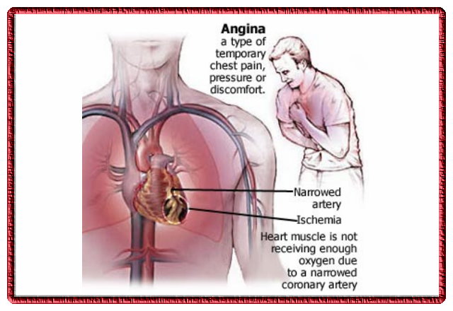
A.pectoris-
 Transient myocardial ischemia.
 It is dueto
1) obstruction of coronary blood flow by atheroma.

2) coronary arterial spasm.

Factors worsen angina-
1 exercise
2 anemia
3 tachycardia
4 HTN


5 hyperthyroidism
6 aortic stenosis
7 aortic reguqitation
8 arrythmias

      C/F -
 History-
1 Retrosternal pain brought on exertion. Relived by rest n sublingual nitrates. It is squeezing,aching. Pain radiates to left arm.
2 angina decubitus is pain while lying flat.
3 Nocturnl angina in aortic regurgitation.There is paroxysmal nocturnal anginal pain with nightmares, dyspnoea, palpitation,skin flushing
4 prinzmetls angina pain which becomes capriciously dueto coronary arterial spasm n ST segment elevation.

PHYSICAL EXAM-

1 rise in BP n heart rate.
2 Fourth heart sound
3 Murmur of mitral regurgitation
4 dyskinetic segment around the apex.
5 paradoxial spliting of 2nd heart sound
6 relive of pain by carotid sinus massage.

    INVESTIGATION-
1 ECG -Reverssible ST segment depression or elivation with or without T wave inversion.

2 Myocardial perfusion scaning using radioactive thallium.

3 echo cardiography n radionuclide scaning pool.

4 coronary arteriography

5 intra vascular ultra sound

     MANAGEMENT-
  3 phases
1 assesment of severity of disease n extent
2 measures to control symptoms
3 measures to improve life expectancy.

GENERAL
1 proper explanation abt disease.
2 avoid walking after meals,in cold.Against wimd.
3 hyperlipidemia treated with drugs n diet
4 control of HTN n DM

DRUGS
1 nitrtes
2 beta adreno receptor antagonist.
3 calcium antgonist
4 plateltes inhibitors.

Nitrates-
glyceryl nitrates(500micro gm) sublingualy.
GTN acts by arteriolar n venous dilatation.
ISOSORBIDE  DINITRATE(10-20mg) three-6 times day.
ISOSORBIDE MONONITRATE.

BETA BLOCKERS-
they reduce myocardial O2 demand by reducing hert rate.
PROPRANOLOL(20mg/3times day)
cardioselective METOPROLOL n ATENELOL.

Ca antagonist-
NIFEDIPIN
VERAPAMIL(40-80mg/3time)

ANTIPLATE agents-
ASPIRIN(75-150mg/day)
CLPIDOGREL

   SURGICAL
 CORONARY ARERY BYPASS GRAFTING.-length of pts saphenous vein is anastomoses to aorta at one end n coronary vessels distal to stenosis to other.
PERCUTANEOUS CORONARY Intervanation.-
(PTCA)percutaneous transluminal coronary angioplasty dilatation of coronary artery stenosis by a small balloon introduced percutaneously via an arterial catheter.

August 12, 2016
Angina pectoris
Angina pectoris


Angina pectoris

*it is a clinical syndrome of discomfort due to transient myocardial ischaemia
*transient ischaemia is due to
1) obstruction of coronary flow by atheroma
2) coronary arterial spasm
3) risk factors -exercise, hypertension, AS, AR, hyperthyroidism
Clinical features
1- retrostermal pain which is squeezing crushing in character. It


commonly radiates to left arm
2- Angina decubitus is a pain while lying flat
3- Nocturnal angina that occurs in aortic regurgitation which is characterised by paroxysmal nocturnal pain associated with nightmare dyspnea palpitation profuse sweating
4- Sprinz metal angina or variant angina is pain which comes capriciously due to coronary artery spasm &accompanied by transient ST segment elevation
5- on examination -*rise in blood pressure & heart rate *fourth heart sound *pansystolic murmur *paroxysmal splithng of second heart sound *relief of pain by cardiac sinus massage (Levine test )
 MANAGEMENT
A) INVESTIGATIONS
*ECG- reversible ST segment elevation /depression with or without T wave inversion
*Echocardiography
*coronary coronary angiography
*CT&MRI are investigation  of choice
B) TREATMENT
*Aims of managements involves 3 phases
1- assesment of severity &extent of disease
2- measure to control symptoms
3- measure to improve life expectancy
*specto discribed under 3 headings
1) General measures
2)Drug treatment
3)Surgical treatment
GENERAL MEASURES
*proper counselling
*avoid walking after meal in cold, against wind
*control hypertension diabetes
*correction of ppt factors
DRUG TREATMENT
1)Nitrates- glycerol trinitrate 500micro gm sublingual relieve pain
2) Beta blockers- they reduce myocardial oxygen demone by reducing heart rate
3) Calcium blockers indicated in if response to beta blockers inadequate / H/o of asthma /prinz metal's angina/sick sinus syndrome
4)Antiplatelet agents low dose aspirin 75 to 150 mg /day
SURGICAL TREATMENT
1)coronary artery bypass grafting by saphenous vein
2)percutaneous transluminal coronary angioplasty 

August 11, 2016
Aneurysm
Aneurysm


Aneurysm

DEF
.
Its a localired or diffuse dilatation of an artery.

TYPES.

1. True: It contains all 3 layers of artery wall.
2. False: it has a pinke layer of fibrous tissue as the wall of the sac and does not contain the three layers of the artery wall as the


covering
majority of false types follow trauma. A mycotic aneurysm which is produced by growth of micro organisms in the vessel wall is of false variety.
3. Arteriovenous: It is a communication betn an artery and adjacent vein.

CAUSES.

Except traumatic all other aneurysms are caused by weakening of the wall of artery.
A]CONGENITAL
1. In circle of willis due to congenital efficiency of elastic lamina at d river of branching. This is called as berry aneurysm.
2. A crisoid aneurysm is the mass of dilated pulsating serpiginous vessels
3. Congenital arteriovenous fistula
4. It is associted with some inherited connective tissue disorder like Marfan and Ehler-Danlos
5. Proximal to coarctation of aorta.
6. Congenital intrimomedial mucoid degeneration.

B]ACQUIRED.
1. Traumatic.
Eg. Direct trauma such as penetrating wound, irradiation or indirect trauma.
2. Degenerative.
This is d most commonest cause majority of them caused by atherosclerosis
3. Infective..
Syphilis, acute infections, mycotic aneurysm, infective endocarditis, tb, arteritis
4. Iatrogenic as in renal dialysis for AV type.

It can also be classified according to shape.
A] Fusiform. It is d commonest variety. It is a spindle shared enlargement.
B] Saccular. It is an expansion of a part of circumference of the artery wall. Usually traumatic.
C] Dissecting. It occurs when the initima ruptures usually beneath an atheromatus plaque & blood is forced through d intima to enter betn inner and outer coats of tunica media.

C/F.

1. May be asymptomatic.
2. Commonest presentation is with dull aching pain.
3. Pulsatile mass
4. severe ischemia of lower limb.
5. Enlargement of artery may block the veins by direct pressure or may cause it to thrombose.
6. severe AV type, may precipitate CCF.
7. Increased limb length in AV type.
8. In AV type below d fistula there may muscle wasting, ulcer formation & cold temp.

O/E
1. Expansile pulsation of d swelling.
2. Pulsation diminishes if pressure is applied proximal to d swelling. It refills again in few beats if pressure is released.
3. Swelling is compressible.
4. Thrill may palpable
5. Systolic bruit
6. Branham's sign:(for AV type)
If a finger is pressed on the artery proximal to the fistula there will be slowing of pulse rate and rise in d diastolic pressure

COMPLICATIONS.

1. Pressure on adjacent structures.
2. Thrombosis & emboli formation.
3. Infection.
4. Rupture.
5. Spontaneous cure.

D/D.

1. Swelling over an artery
2. swelling beneath an artery
3. Pulsating tumor
4. Abscess.

INVESTIGATIONS.

1. Blood is examined for cholesterol levels.
2. W.R. & Kahn tests to exclude syphilis.
3. Straight x-ray.
4. Arteriography
5. Investigations for anesthetic & surgical fitness.

TREATMENT.

1. Arterial Ligation:
suitable for splenic a. or intracranial aneurysm.
#Anel's- applied just proximal to sac
#Brasdor's- applied just distal to sac.
#Hunter's- applied immediately above a branch of artery.
#Wardrop's- applied immediately below a branch of artery.
#Antylus's- applied one proximal & one distal to sac.

2. Wiring of aneurysm sac.
Indicated in elderly and poor risk patients and in difficulty placed aneurysm.

3. Wrapping of aneurysm sac.
Used in intracranial aneurysms.
A strip of fascia lata, polytiene or cellophane is wrapped round d aneurysm to strengthen its wall

4. Mata's aneurysmorrhaphy.
Suitable in femoral or popliteal artery.
Sac is totally excised & defect in d wall is closed by suturing adjacent healthy artery wall.

5. Exclusion & bypass grafting.
Used where excision of sac is not safe due to adhesions with neighbouring vital structures.

6. Excision and grafting.
Most popular & should be done wherever possible. Dacron graft or autogenous vein is used.

7 .Excision and end to end suturing.
Possible only in cases of peripheral aneurysm. Two cut ends are approximated..

8. For AV type:
# reconstructive operation is the treatment of choice
# Quadruple ligation
# selective intra-arterial embolisation
# amputation of limb in cases of severe ischaemia.

August 10, 2016
Anorexia nervosa
Anorexia nervosa

Anorexia nervosa

There is marked weight loss arising from food avoidance. often c- purging bingeing . exce
exercise or the use of diuretcs&
laxatives.
Patient Still feel overweight
false beliefs r delusional
Anxiety n depression.
Downy hairs.
ETIOLGY
unknown but probably
enviorn n genetisc factors
social pressure on women.


PHYSICAL CONSEQ
CARDIAC T wave inversion ,ST depression ,arrythmia
HAEMATOLOGICAL
anemia, thrmbocyto,leucopenia.
ENDOCRINE pubertal delay ,short stature ,amenorrhoea
GIT constipatin .abn LFT.
DIAG CRIT
wt loss at least 15%
avoidance f high calorie foods.
Distortion f body image.
Amenorrhoea for at least 3 months.
MANAGEMENT
Ensure Patients well being
Psychological traument CBT N family therapy.

August 09, 2016
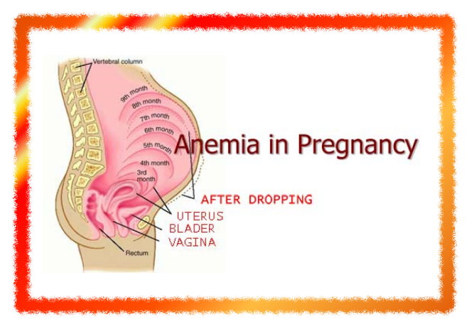
Anaemia in pregnancy


Anaemia in pregnancy

Classification of anamia:
1) physiological anaemia of prg.
2) pathological
 A) iron deficiency anaemia
     iron, folic acid, vit B 12 and protein def
 B) haemorrhagic anaemia
        a) acute - following bleedin in early months
        b) chronic - hookworm infestation and


           bleeding piles
 C) heriditary
        a) thalacemia
        b) sickle cell anaemia
 D) bone marrow insufficiancy
 E) ananemia of infection
 F) chr disease or neoplasm

Aetiology of iron deficiency anaemia
 - low iron intake
 - increase nutritional demand
 - poor absorption and utilisation
 - poor reserve
 - increased loss of iron

C/F
 A) symptoms
    - fatigue, weakness, lassitude
    - impaired work capacity
    - dizziness, headache insomnia
    - ankle oedema
    - dyspnoea, palpitaion
    - anorexia
 B) signs
    - pallar of skina nd mucus memb in severe
      anaemia
    - loss of palmer creases
    - glossitis, dysphagia, stomatitis
    - koilnychia
    - tachycardia

Effect of anaemia on pregnancy
 A) mother
    1) during pregnancy
       - cardia failure
       - increase susseptibility of infection
       - preterm labour
       - pre-eclampsia
    2) during labour
       - uterina inertia
       - PPH
       - cardiac failure
       - shock
    3) during puerperium
       - cardiac failure
       - puerperial sepsis
       - failing lactation
       - chronic ill health, back ache

 B) fetus and neonate
    1) prematurity
    2) IUGR and death

Investigations
 A) for degree of anaemia
   1) hb%
      Mild 8-10 gm%
      Moderate 7-8 gm%
      Severe less than 7%
   2) rbc count usually less tham 4 millions/ml
 B) to know type of anaemia
   1) peripheral smear
     - to know marphology of RBC
     - mocrocytic hypochromic
   2) haematological indices
     - MCH less than 25 pg
     - PCV less than 30%
     - MCHC less than 30%
     - MCV less than 75 μmc
 C) stool examination
    - to detect parasitic inf
       Like, ankylostoma, hookworm,
 D) urine examination
    - for presence of protein, sugar and pus cells
 E) specific investigations
    - vit B12 and folic acid in case of megaloblastic
      anaemia
 E) other blood values
    - serum iron less than 30 μg/dl
    - TIBC more than 400 μg/dl
    - percentage saturaion 10% or less
    - serum ferritin less than 15μg/lit

Treatment
 (A) general prevention
     - avoidance of frequent child births, minm interval shud b atleast 2 yrs
     - supplimentary iron therapy- ferrus sulphate 200 mg (60 mg of elemental iron) with 1 mg of folic acid
     - dietary prescription
          Diet rich in iron and prtein like liver, vegitables, meat, eggs, jaggery
     - fortification of food and salt by iron
     - adequate rx of hookworm, bleeding piles, malaria, dysentru, UTI
     - screening of adolescence girls in school

 B) definative rx
   1) iron therapy
      a) oral iron therapy
         - indicated in mild to mod anaemia
         - 100-200 mg elemental iron per day
         - common preparations r ferrous sulphate, ferrous fumarate, ferrous fumarate
         - innitial dose- 200 mg ferrous sulphate thrice daily
         - if large dose is required, shuld b raised gradulay in 3-4 days
         - rx shuld b continued till blood picture bcomes normal
        - thereafter maintainance dose of 1 tab daily for atleast 100 days following delivery to replenish iron stores
        - side effects- nausea, vomiting, intolerance, diarrhoea, constipation, epigastric pain
      b) parentaral iron therapy
        Indications
         - intolarance, noncompliace, poor absorption of oral therapy
        Preparatio
        - iron sorbitol citric acid complex
        - iron dextran
        Given im 100 mg/day on alternate day by Z technique
        Side effects- painfull, abcess and staining at inj site
       - intavenous route, required amt of iron (mg) =25 mg*percentage deficit
        Complications r thrombophlebitis, fever and malise
     Oral iron shuld be stopped b4 40 hr of parentaral iron
 
   2) blood transfusion
       - svr anaemia, svr haemorrhage ( aph, pph, cs), thalasemis, sickling dissorder
                               - by mak and sushil

August 09, 2016
Analgesic nephoropathy

Analgesic Nephoropathy

 Heavy use of amalgesic mixture of phenacetin in combination with aspirin acetaaminophen
Morphology
 papillary necrosis
 tubulo interstetial infalmation
damage to vascular supply of inner medulla lead to local intersti inflam reaction
 
c/f          
proteinuria
hematuria
pyuria
renal coli
nephrocalcinosis

invest

urine -proteinuria
     hematuria
     pyuria

IPV
 ring sign

CT
 gasRland paTERN

TREATMENT
discourage use of this combinatio n phenactin              

August 09, 2016
Astigmatism
Astigmatism

Astigmatism

Astigmatism is type of refractive eror in which refraction varies in diffrent media.
Types-regular and irregular.
Regular=refractiue power changes uniformly frm one meridian to another.
ETIOLOGY-1.Corneal astigmatism is d result of abnormalities of curvature of cornea.Most comon.
2.Lenticular astigmatism-a)curvatural due to abnormalities of curvature of lens.
b)positional due to tilting of lens as in subluxation..
c)index astigmatisms may ocur rarely.
Types of regular astigmatism-1.With rule astigmatism=when 2 principal meridia r placd at right angles to one another but vertical meredian is more curvd than horizontal.Corection requires cocave cylindes at 180 +-20 degree or convex cylindrical at 90+-20
2.Against rule astigmatism refers to condition in which horizontal meridian is more curvd than vertical.Correction requires convey cylindrical lens at 180+-20degree.
3.Oblique astigmatism-in which two principal media s nt horizontal and vertical thougi these are at right angle to 1 another.Corection requires cylindrical lens at 30 in both eyes.Or comlimentary cylindrical in 30 in 1 eye and 150 in other eye.
4.Bioblique astigmatism-two principal media r nt at right angle to each other..
Refractive type of regular astigmatism-1.Simple when rays r focussd in one meridian and either in front or behind. Simle myopic or simple hypermetropic respectively.
2.Compound-in which rays of light in both the meridia r focusd either in front or behind.
3.Mixed astigmatism in which light rays in 1 meridian r focusd in front and in other meridian behind the retina.Thus 1 meridian eye is myopic and another hypermetropic.
Symptoms-1.Defective vision 2.Blurin of objects .3.Object may b proportionaly elongated4.Asthenopic symptoms.
Signs-1.Difrent power in 2 media.
2.Oval or tilted disc
3.Head tilt-may develop torticolis to have good vision.
4.Half closure of eyelid.
INVESTIGATIONS-
1.Retinoscopy reveal difrent power in 2 difrent axis
2.Keratometry
3.Astigmatic fan test and jackson cross cylinder test.
TREATMENT-1.Optical treatment of regular astigmatism comprises following
a)spectacle with ful corection of cylindrical power and corect axis.
b)contact lenses-rigid contact lenses may corect upto 2-3 of regular astigmatism
2.Surgical correction of astigmatism is quite efective.
IRREGULAR ASTIGMATISM-
Characterisd by an irregular change of refractive power in diffrent media.
Etiological types-
1.Curvatural irregular astigmatis m found in patient of extensive corneal scar.
2.Index irregular astigmatirm due to variable refractive index in difrent parts in lens.
Symptoms-1.Defective vision
2.Distortion of objects
3.Polyopia
Investigations-1.Placido disc test reveals distorted circles.
2.Photokeratoscopy and computerisd corneal topography reveal irregular corneal curvature.
TREATMENT-1.Optical treatment consists of contact lens which replaces anterior surface of cornea.
2.Phototherapeutic keratectomy may b helpful.
3.Surgical treatment indicated in extensive corneal scarring and consists of penetrating keratoplasty.

August 08, 2016
Atrial fibrillation
Atrial fibrillation


Atrial Fibrillation

Definition:
 This is an arrhythmia where atria are disorganised and multiple atrial foci fire impulses at a rate of 350-600 per minute. There is no atrial contraction but only fibrillation. The ventricles respond at irregular intervals, usually at a rate of 100-140/min.
 It can be paroxysmal or persistent.

Aetiology:
1. Rheumatic heart disease.
2. Ischaemic heart disease.


3. Hypertension.
4. Thyrotoxicosis.
5. Congenital heart diseases.
6. Cardiomyopathy.
7. Pericardial diseases.
8. Lone atrial fibrillation occurs in elderly patients without underlying heart disease.

Pathophysiology and haemodynamics.
1. Atria fire the impulse at a rate of 350-600/min. Many of them reach the AV node in the refractory period, and so not conducted. However, a variable number of impulses are conducted to the ventricles at irregular intervals. This accounts for the irregularly irregular rhythm of pulse and heart.
2. The irregular rhythm of heart results in varying durations of diastole, the lesser the ventric volume and the longer the diastole, the higher the ventricular volume. The ventricular volume in turn determines the cardiac output, which hence keeps varying. This varying cardia output accounts for the warying volume of pulse deficit.
3. Lack of atrial contraction results in the following.
a. Stasis of Blood in the left burgun resulting in thronat formation and subsequent dislodgement of the thronat resulting in sustemic embolisation:)
b. Disappearance of a wave from JVP.
c. Disappearance of fourth heart sound he it was present.
d. Disappearance of the pre-systolic accentuation of the mid-diastolic murmer of mitral stenosis in some cases.
4. Tachycardia-related cardiomyopathy in patients with poor rate control may further depress cardiac function.

Risk of stroke in atrial fibrillation.
CHADS2 index. It has following components. The numerals in front of the components indicate the score.
 Congestive heart failure   1
 hypertension                 1
 age more than 75 years   1
 diabetes mellitus             1
 stroke history               2

score of 6 predicts a stroke of 18.2 per 100 patient-years.

Symptoms
1.palpations
2.fatigue
3. Syncope
4. Angina
5. Symptoms of cardia failure and thromboembolism.

Signs.
 Irregularly irregular pulse.
 Varying volumes of pulse.
 Pulse deficit.
 Varying intensity of first heart sound.
 Absence of a water onJVP
 disappearance of the  PSA of the MDA of MS in some cases.
Disappearance of the fourth heart sound.
 Hypottension

electrocardiogram
. An irregularly irregular rhythm of QRS complexes.
. Absent P wave.
. Small, irregular wave at a rate of 350-600/min .

Differential diagnosis.
. Atrial flutter with variable block. Prominent saw-tooth waves at lower rates of 250-350/min are seen in the ECG.
. Atrial tachycardia with vagable block. Atrial rate 150/min approximately which is regular but the and to the wentsic is not regular producing irregular pulse.
. Multifocal burial tachycardia.

Complications.
.Syncope
. Thromboembolism
. Precipitation of cardiac failure.
.Angina
. Hypottension.
. Precipitation of pulmonary offenc in nitrat stenosis.

Treatment.
Goals of management.
. Haemodynamic stabilization.
.Control of ventricular rate.
. Restoration of shots rhythm.
. Prevention of emboli complications.
. Treatment of underlying cause.
1. If the patient 's clinical status is not severely compromised, synchronized DC cardioversion starting at 100J is the treatment of choice.

2. He the patients clinical status is not peter compromised, treatment is in two steps :
a. Slowing the ventricular rate with verapamil, diltiazem, propranolol, esmolol or digoxin. The doses of various drugs are.
Diltiazem:10mg intravenously over 2 min, repeat same ford in15 minutes if required ; start an infusion at 10-15 mg/hr to maintain ventricular rate below 100/minute.
Digoxin:0.25 -0.5 mg intravenously, then 0.25 mg after 4-6 hours joe another ford after another 12 hours.

b. Converting rhythm to normal shots rhythm.
 Amiodarone 5-7 mg/kg iv over 1 hour followed by 1.2-1.8gm/24 hour infusion.
If medical cardioversion fails, electric cardioversion is performed after 3 weeks of warfarin therapy which is continued for another 4 weeks after cardioversion.

3.Quinidine or amiodarone may be used to prevent recurrence.
4. If cardioversion is unsuccessful or in which it is likely to reccur, allow the patient to remain in atrial fibrillation but reduce the ventricular rate by digitalis, verapamil or propranolol. Or give chronic anticoagulation
5. Antithromantic therapy is indicated in the following situations:
a. Duration of atrial fibrillation exceeds 48 hours
b. If atrial thrombi are seen on echocardiography. Warfarin is administered for 3 weeks prior to cardioversion. It is continued for another 4 weeks after cardioversion.

6. In paroxysmal atrial fibrillation., preservation of shots rhythm can be achieved quinidine, amiodarone.
7. Refractory cases managed with antitachycardia racemclers or inducing complete heart block by ablation of bundle of His followed by permanent pacemaker implantation.

MKRdezign

{facebook#https://www.facebook.com/suredevelopersteam/} {twitter#https://twitter.com/sskhanontoday} {google-plus#https://plus.google.com/+SskhanontodaySureshahebazkhan} {pinterest#https://in.pinterest.com/sskhanontoday/} {youtube#https://www.youtube.com/channel/UCHvWgxgeLRtrfInYzn1PcHg/featured?view_as=public} {instagram#https://www.instagram.com/sskhanontoday/}

Contact Form

Name

Email *

Message *

Powered by Blogger.
Javascript DisablePlease Enable Javascript To See All Widget